Does Blood Turn Red in the Air? The Complete Science Guide to Blood Color and Oxygen
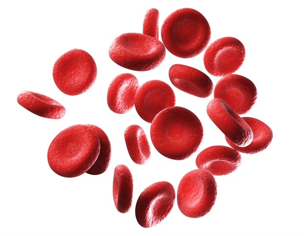
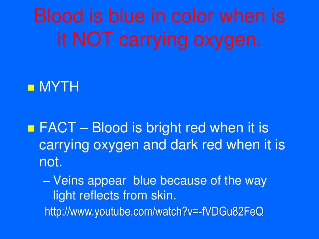
Millions of schoolchildren grew up hearing a persistent anatomical rumor: human blood runs blue through your veins until an open wound exposes it to oxygen in the ambient air. It feels intuitive when looking down at the pale undersides of your wrists, where faint, turquoise-tinted vessels spider under the skin. Yet that belief remains entirely false. As detailed in a News Medical Report covering breakthroughs in microvasculature imaging, advanced optical platforms now track 3D erythrocyte oxygen transport down to the single-cell level inside intact living tissue. The physics and biology are unambiguous: human blood is never blue at any point in the circulatory system.
Whether coursing through high-pressure arteries straight out of the lungs or creeping back to the heart through low-pressure systemic veins, human blood stays red. The precise hue fluctuates depending on chemical binding, but it never crosses the spectrum into azure or cobalt. Understanding why this myth survives requires unpacking both the biochemistry of hemoglobin and the deceptive optical properties of living human tissue.
📌 Key Takeaways:
- The Direct Fact: Human blood is never blue inside the body; it ranges from bright scarlet when oxygen-rich to a deep, dark maroon when depleted.
- The Underlying Cause: Veins appear blue exclusively due to optical scattering, red light wavelengths penetrate deep into tissue to be absorbed, while high-energy blue wavelengths reflect off skin layers.
- True Blue in Nature: True blue blood exists only in invertebrates like horseshoe crabs and octopuses, which rely on copper-based hemocyanin rather than iron-based hemoglobin.
The Dark Red Reality of Deoxygenated Blood
The human circulatory system relies on red blood cells packed with hemoglobin, a complex protein made of four globular subunits. Each subunit cradles a heme group centered around an iron ion ($Fe^{2+}$). This iron binding architecture dictates the liquid's interaction with ambient light.
When blood flows through the pulmonary capillaries, oxygen molecules bind directly to these iron sites. This binding event physically twists the hemoglobin protein, shifting its tertiary structure into what biophysicists call the "relaxed" state. In this configuration, the molecule absorbs blue-green light wavelengths while reflecting long, vibrant red wavelengths back to an observer. The result is oxygenated blood: the bright, cherry-scarlet fluid that sprays from an arterial puncture.
After red blood cells navigate the body's capillary beds and surrender their oxygen payload to metabolic tissues, hemoglobin snaps into a "tense" deoxygenated conformation. This structural shift alters the light-absorption spectrum. Deoxygenated blood does not turn clear, nor does it morph into blue. It darkens into a dense, purplish maroon.
Phlebotomists witness this color during routine clinical blood draws. When an evacuated needle enters a cubital vein, the vacuum tube contains zero ambient air. The fluid surging into the vial never touches atmospheric oxygen. It immediately appears as a dark, murky crimson, irrefutable physical evidence against the dark red blood myth.
Skin Optics and Why Veins Look Blue to the Human Eye
If venous blood is dark maroon, why do superficial veins look visibly blue or green through the skin? The answer is not hematology; it is physics.
The color human eyes perceive when looking at a superficial vein is an optical illusion created by light wavelength penetration across multiple dermal layers. White light from the sun or a light bulb comprises the entire visible spectrum, ranging from short, high-energy blue wavelengths (around 400 to 475 nanometers) to long, low-energy red wavelengths (around 620 to 750 nanometers).
Human tissue, subcutaneous fat, and skin do not handle these wavelengths equally:
- Red wavelengths: Possess deeper penetration power. They push deep through the epidermis and dermis. Because deoxyhemoglobin absorbs red light efficiently, the vein beneath the skin absorbs these penetrating red rays. They do not bounce back to the eye.
- Blue wavelengths: Lack the energy penetration to pass deep into tissue. Instead, they hit the upper dermal matrix and scatter almost immediately, an effect closely related to Rayleigh scattering.
- The Perceptual Contrast: The skin immediately adjacent to the vein reflects both red and blue light back to your eyes. The skin directly overlying the vein reflects mostly scattered blue light, because the red light was absorbed by the underlying vessel.
Your brain compares the light coming from the skin above the vein with the light coming from surrounding tissue. Because the vessel selectively absorbs red rays while the surface tissue scatters blue rays back, the brain processes the relative difference as a cool blue or green stripe. In people with thinner skin or less melanin, this chromatic contrast is even more pronounced.
Comparative Hematology: Which Animals Actually Bleed Blue?
While human blood never turns blue, the animal kingdom features diverse respiratory pigments tailored to extreme environments. The color of an organism's circulatory fluid depends entirely on the metal ion anchoring its primary transport protein.
| Respiratory Pigment | Active Metal Core | Oxygenated vs. Deoxygenated | Key Animal Groups |
|---|---|---|---|
| Hemoglobin | Iron ($Fe^{2+}$) | Bright Scarlet / Dark Maroon | Humans, mammals, reptiles, birds, fish |
| Hemocyanin | Copper ($Cu^{2+}$) | Vivid Cyan Blue / Colorless Clear | Horseshoe crabs, octopuses, squids, snails |
| Chlorocruorin | Iron ($Fe^{2+}$) | Dull Green / Light Brownish | Certain marine polychaete annelids |
| Hemerythrin | Iron ($Fe^{2+}$ / $Fe^{3+}$) | Violet-Pink / Colorless Clear | Brachiopods, peanut worms, priapulids |
Invertebrates such as the Atlantic horseshoe crab (Limulus polyphemus) and cephalopods do not produce hemoglobin. Living in cold, low-oxygen marine environments, they rely on hemocyanin. Rather than floating encapsulated inside distinct red cells, hemocyanin molecules float freely throughout the animal's hemolymph.
Copper drives this reaction. When oxygen binds between two copper atoms, electronic transitions generate strong light absorption in the orange-red spectrum, leaving behind an intense sky-blue color. When an octopus uses up its oxygen supply, its blood turns entirely clear and colorless.
Real-Time Single-Cell Imaging Erases the Last Ambiguities
For decades, proving that blood never turns blue inside deep internal organs relied on indirect blood draws or invasive surgical observation. Skeptics frequently argued that cutting through tissue exposed internal blood vessels to ambient atmospheric air or harsh operating room lamps, artificially altering the fluid's appearance before human observers could view it.
Technological advances in high-speed microvasculature imaging have permanently eliminated that counter-argument. Modern biomedical laboratories deploy non-invasive two-photon intravital microscopes capable of imaging living animal and human capillaries through intact dermal windows.
These optical systems capture erythrocyte oxygen transport at single-cell resolution. Researchers monitor individual red blood cells as they squeeze single-file through capillary loops measuring under 8 microns across.
By measuring the intrinsic phosphorescence decay and autofluorescence of native hemoglobin, researchers map exact oxygen saturation gradients across cerebral and dermal tissues. The imaging confirms that deoxygenated erythrocytes maintain an uninterrupted, dark red absorption signature even in hypoxic niches where oxygen pressure approaches zero. There is no intermediate blue transition phase.
How Medical Illustration Reinforced a Cultural Fallacy
If physiological data has disproven the myth for over a century, why do people still believe it? The blame rests largely on pedagogical convention in early education.
Standard anatomy textbooks, classroom posters, and medical diagrams use color-coding to clarify complex circulation paths:
- Red Lines: Signify oxygen-rich systemic arteries carrying blood away from the left ventricle.
- Blue Lines: Signify oxygen-depleted systemic veins returning blood toward the right atrium.
Medical illustrators trace this visual convention back to the pioneering anatomical drawings of the 16th and 17th centuries, codified systematically by publishers in the 19th century. Using contrasting primary colors solved a layout problem: black-and-white etchings made it impossible to distinguish intertwined vascular networks running in parallel across the extremities.
Generations of elementary and middle school students absorbed these simplified visual aids literally. Lacking training in graphic design conventions, young minds concluded that the human body runs on two distinct colored fluids: bright red juice in the arteries and blue ink in the veins. When paired with the visible blue appearance of surface wrist veins, the misconception hardened into popular folklore.
Frequently Asked Questions (FAQ)
Q1: Why does venous blood look almost black when drawn in large volumes?
A1: Deoxygenated blood is a deep, saturated maroon. When packed densely inside a standard glass collection vial or blood donation bag, the high concentration of light-absorbing deoxyhemoglobin blocks light transmission entirely. The lack of reflected light makes the volume appear black or muddy brown to the naked eye.
Q2: Can any rare medical disorder actually turn human blood blue?
A2: No condition turns human blood blue, though some make blood look chocolate brown. In methemoglobinemia, the iron in hemoglobin oxidizes from $Fe^{2+}$ to $Fe^{3+}$, preventing oxygen release to tissues. This turns the blood dark brown and starves skin of oxygen, causing severe cyanosis, a condition where lips and fingers appear pale blue, even though the blood itself is brown.
Q3: Does atmospheric oxygen instantly brighten spilled blood?
A3: Yes. When venous blood leaves the body through a cut, dark maroon deoxyhemoglobin binds immediately to ambient atmospheric oxygen on the fluid's surface layer. This rapid surface re-oxygenation shifts the molecular conformation, turning the spilled droplet bright scarlet within milliseconds.
The Structural Reality of Human Circulation
Human blood operates on a tightly conserved iron-based transport system that remains crimson across every phase of life. From the moment red blood cells pick up oxygen in the alveolar capillaries to the second they return through systemic veins, their structural palette never touches blue. The turquoise vessels branching across your wrists are an optical mirage born from light scattering through living skin, while the blue arrows of childhood biology charts are nothing more than helpful graphic design. Authentic blue blood belongs strictly to the deep sea, leaving human bodies anchored firmly in the red.
