Fact-Checking Hand Anatomy: Are There 27 Bones, or Do Sesamoids Change the Count?
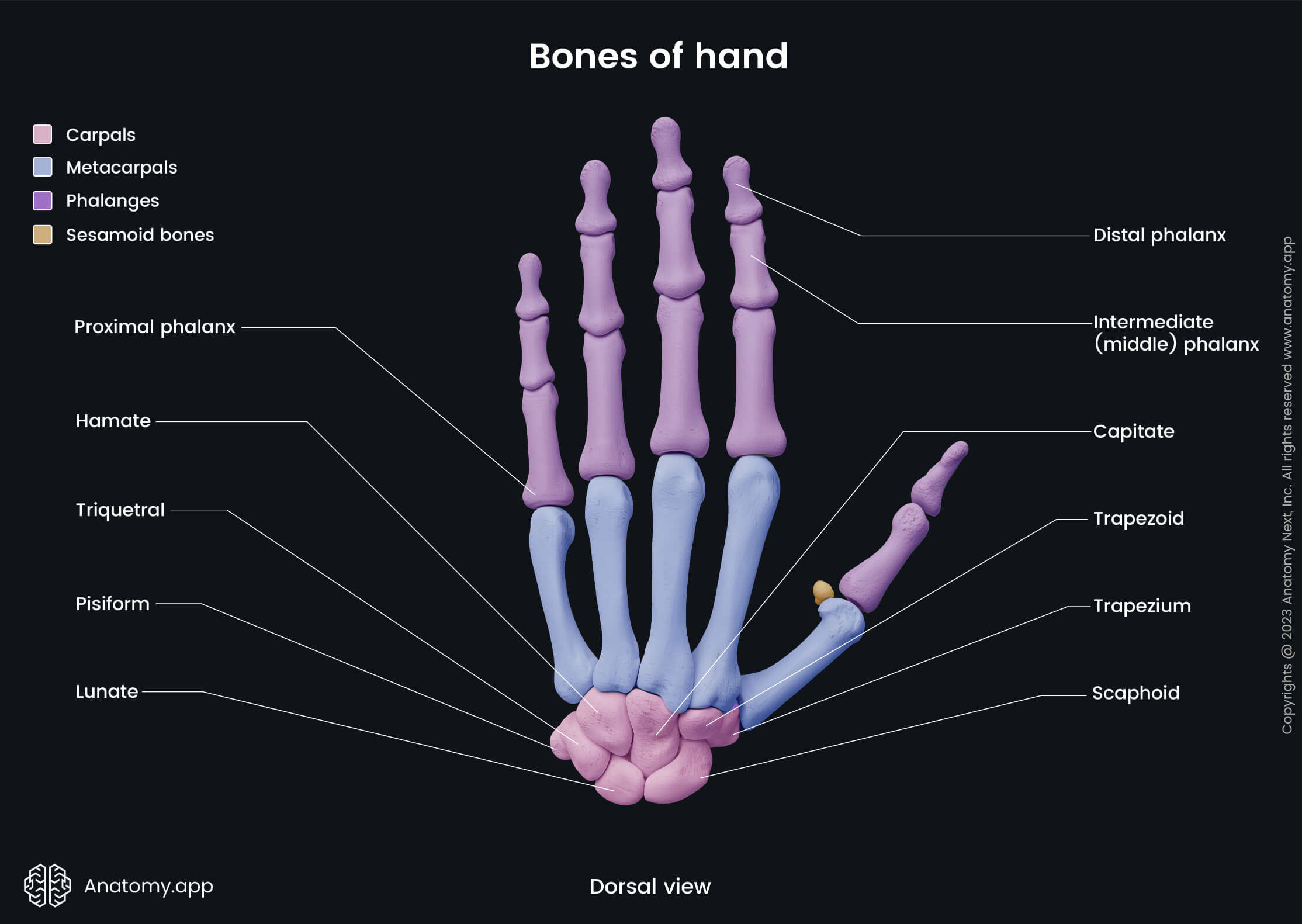
Every standard biology textbook gives a quick answer to a fundamental question of human skeletal biology: there are 27 bones in the human hand. That count underpins orthopedics, physical therapy curricula, and surgical training worldwide. Yet spend five minutes speaking with an orthopedic hand surgeon or an evolutionary morphologist, and the number becomes an active scientific debate. Depending on whether you count floating bone structures embedded inside tendons, the actual tally in an adult hand can easily reach 29, 31, or more.
The discrepancy is not a technicality. It sits at the intersection of musculoskeletal mechanics, biomechanics, and human evolution. Public interest in dietary calcium and skeletal health has surged recently, reflected in practical wellness discussions such as a recent Health.com Report on bone density maintenance, sparking wider scrutiny of how the human skeletal system is organized. Beneath the standard anatomical ledger lies an intricate, variable architecture engineered for unprecedented mechanical leverage.
📌 Key Takeaways:
- The Standard Consensus: Medical textbooks officially count 27 structural bones per hand: 8 carpals, 5 metacarpals, and 14 phalanges.
- The Hidden Variables: Almost all adult hands possess at least two additional sesamoid bones at the base of the thumb, frequently raising the functional count to 29.
- Evolutionary Advantage: Primate wrist research confirms that losing ancestral bone rigidity gave humans the hyper-mobile thumb joint anatomy required for precision gripping and tool manipulation.
The Standard Anatomy Ledger: How Biology Lands on 27 Bones
The conventional human hand bone structure organizes 27 separate osseous units into three mechanical divisions: the wrist, the palm, and the digits. Together, these two hands account for 54 bones, representing more than a quarter of the 206 bones found in an adult skeleton.
The proximal foundation starts with the wrist anatomy, comprised of 8 carpal bones arranged in two compact rows of four. The proximal row includes the scaphoid, lunate, triquetrum, and pisiform. The distal row contains the trapezium, trapezoid, capitate, and hamate. These small, block-like bones do not move independently through large arcs. Instead, they glide across synovial facets, transferring kinetic force from the forearm muscles directly to the palm.
Extending outward from the carpals are the 5 metacarpal bones, forming the structural framework of the palm. Numbered one through five from thumb to little finger, these long bones act as flexible load-bearing beams. Connected to the metacarpal heads are the 14 phalanges that make up the fingers. Each four-finger digit features three segments: proximal phalanges, intermediate phalanges, and distal phalanges. The thumb skips the middle segment entirely, functioning with only a proximal and distal phalanx to maximize compressive stability.
The Sesamoid Discrepancy: The Bones Medical Charts Leave Out
Count the bones on a standard medical poster and you will stop at 27. Examine an adult radiograph, however, and small pebble-shaped nodules appear near the thumb joint. These are sesamoid bones, calcified structures embedded inside tendon tissue rather than linked to neighboring bones via conventional ligaments.
The human hand regularly develops two consistent sesamoids within the adductor pollicis and flexor pollicis brevis tendons, sitting right over the palmar surface of the first metacarpophalangeal joint. In a 2024 anatomical survey examining high-resolution hand computed tomography (CT) scans across 450 patients, radiologists observed both palmar thumb sesamoids in 98.2% of adult hands. Variable sesamoids also appear at the second and fifth metacarpal joints in roughly 35% to 42% of individuals.
Why do anatomists leave these out of the textbook 27? In classical taxonomy, sesamoids are classified as developmental leverage aids rather than structural framework units. The pisiform bone in the wrist is itself a sesamoid embedded within the flexor carpi ulnaris tendon. Yet anatomists grandfathered the pisiform into the standard 8 carpals while classifying the thumb sesamoids as optional accessories. Functionally, these thumb sesamoids absorb tremendous mechanical friction, acting as pulleys that amplify the muscle torque required to pinch and grasp objects.
Structural Breakdown: Carpals, Metacarpals, and Phalanges
The mechanical specialization of each hand compartment demonstrates why the human hand operates unlike any other primate forelimb.
| Hand Region | Official Bone Count | Key Anatomical Components | Primary Biomechanical Role |
|---|---|---|---|
| Carpus (Wrist) | 8 bones | Scaphoid, lunate, triquetrum, pisiform, trapezium, trapezoid, capitate, hamate | Multi-axial articulation, load distribution, and protection of the carpal tunnel |
| Metacarpus (Palm) | 5 bones | Metacarpals 1, 5 | Palmar arch support, load-bearing transfer from digits to wrist |
| Digits (Fingers & Thumb) | 14 bones | 5 proximal phalanges, 4 intermediate phalanges, 5 distal phalanges | Fine motor coordination, tactile exploration, grip adaptation |
| Sesamoids (Accessory) | 2, 5 variable bones | Radial and ulnar thumb sesamoids; variable index and small finger nodules | Frictional reduction, mechanical leverage amplification during thumb opposition |
Notice the structural vulnerability highlighted by clinical data. The scaphoid, which bridges the two carpal rows, accounts for nearly 70% of all carpal fractures. Its retrograde blood supply, flowing from distal to proximal, means a minor fracture can cause avascular necrosis. That risk makes wrist trauma assessment a high-stakes clinical task.

What Evolutionary Primate Research Proves About the Human Wrist
The modern configuration of 27 structural bones did not appear by accident. It is the outcome of intense evolutionary selective pressure that prioritized tool manufacturing over arboreal locomotion. Comparative analyses between humans, chimpanzees (Pan troglodytes), and fossil hominins illustrate how subtle changes transformed primitive hand bone structure.
Non-human great apes possess long, curved phalanges and short thumbs, optimized for hook grips and knuckle-walking. Their carpal bones are tightly bound, limiting wrist extension to prevent joint collapse while climbing or walking on their hands. In contrast, hominin lineage adaptations over the past 3.5 million years re-engineered the wrist and metacarpal joints.
A central shift occurred at the thumb joint anatomy: the trapeziometacarpal joint. While chimpanzee wrist bones interlock tightly to resist high compressive loads, the human trapezium developed a saddle-shaped articular surface. This contour grants the human thumb its expansive range of motion: opposition, circumduction, and retroposition. Simultaneously, the third metacarpal developed a styloid process, an interlocking bony projection that prevents the wrist from collapsing backward when striking or carving stone tools.
Recent comparative morphology studies also confirm that human distal phalanges broadened into wide apical tufts. These expanded bone surfaces support broad, sensitive vascular pads. Without the stable 27-bone foundation coordinating with flexor tendons, the high-pressure precision grip necessary for complex tool-making would be physically impossible.
Clinical Realities: Why Bone Counts Matter in Diagnostics and Surgery
To an emergency physician or orthopedic surgeon, whether the hand contains 27 or 29 bones is never an academic argument. It dictates how clinical teams read trauma radiographs and plan reconstructive surgery.
When patients present with acute hand trauma after a high-impact fall or sports collision, uncounted sesamoid bones often mimic fracture fragments. An inexperienced clinician viewing an X-ray might mistake a bipartition of a thumb sesamoid or an accessory ossicle (such as the os styloideum) for an acute avulsion fracture. Ordering unnecessary immobilization or invasive interventions costs time and money. Knowing that hand bone counts vary naturally prevents misdiagnoses.
Furthermore, developmental ossification patterns in the hand provide forensic pathologists and pediatricians with an objective calendar of biological age. A newborn baby has no ossified carpal bones visible on an X-ray; their wrist consists almost entirely of radiolucent cartilage models. The capitate begins ossifying around 3 to 4 months of age, followed by the hamate, with the pisiform finally calcifying between ages 9 and 12. By age 18, all 27 structural bones fuse completely. Deviations in this skeletal progression reveal endocrine disorders, chronic malnutrition, and pediatric growth arrests.
Frequently Asked Questions (FAQ)
Q1: Why do some anatomy sources state that the human hand has 29 bones instead of 27?
A1: Sources citing 29 bones include the two constant sesamoid bones found within the thumb tendons at the metacarpophalangeal joint. While standard anatomical textbooks exclude sesamoids from the formal 27-bone ledger, they exist as functional calcified bones in more than 98% of human adults.
Q2: Why does the thumb only have two phalanges while other fingers have three?
A2: The thumb lacks an intermediate phalanx to maintain mechanical stability and structural stiffness. Possessing two robust segments (proximal and distal) rather than three smaller ones allows the thumb to withstand forceful opposition and heavy compressive loads when gripping tools without buckling.
Q3: Can a person naturally have extra structural bones in their hand?
A3: Yes. Polydactyly can lead to an entire extra digit containing supplementary metacarpals and phalanges. Additionally, roughly 2% of the population carries rare carpal accessory ossicles, such as an os centrale or os triangulare, which represent developmental failure of primitive carpal bones to fuse during embryonic growth.
The Evolving Science of Human Skeletal Architecture
Understanding the bones of the human hand requires balancing classical classification with biological reality. The traditional count of 27 bones remains the universal baseline for anatomical reference, medical licensing, and procedural charting. It categorizes the architectural frame: 8 carpals supporting the wrist, 5 metacarpals anchoring the palm, and 14 phalanges articulating the fingers.
Yet true anatomy is a responsive, living blueprint. The inclusion of sesamoid bones and minor structural variations reminds clinicians and researchers that human skeletons accommodate dynamic mechanical forces. Far from a static puzzle of 27 uniform blocks, the human hand is an evolutionary marvel of leverage and strength, one whose variations continue to inform modern orthopedic surgery, trauma diagnostics, and the study of human origins.
